Facility
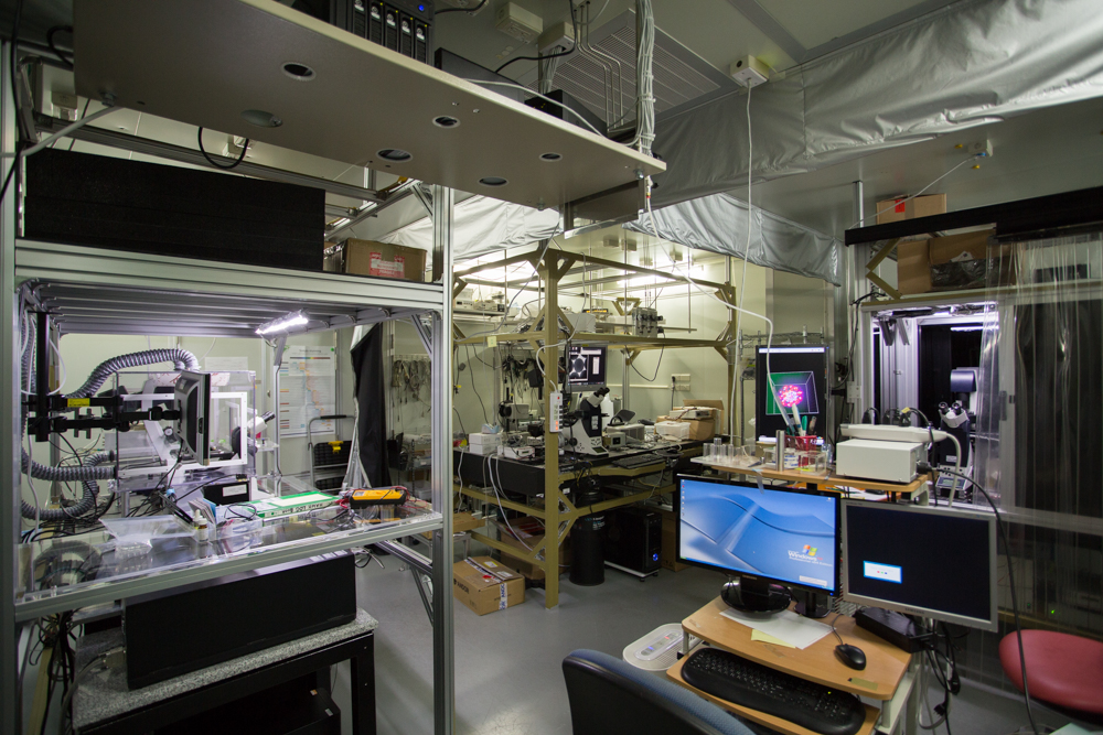
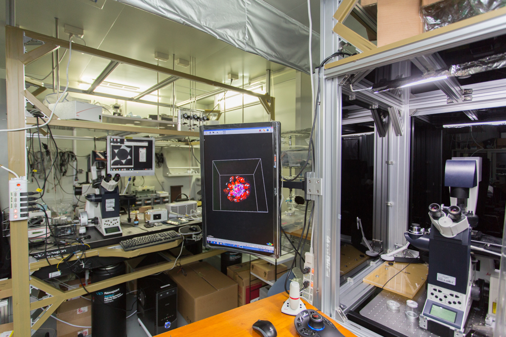
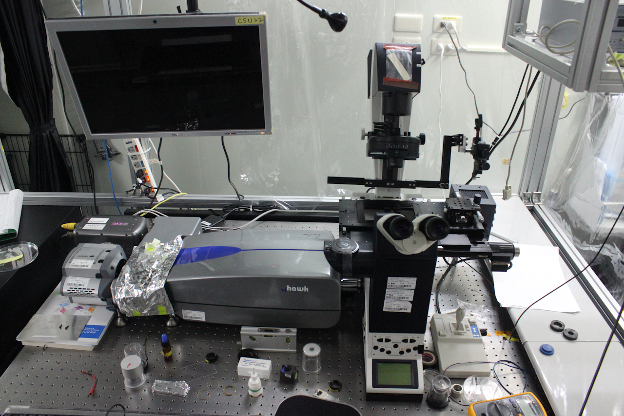
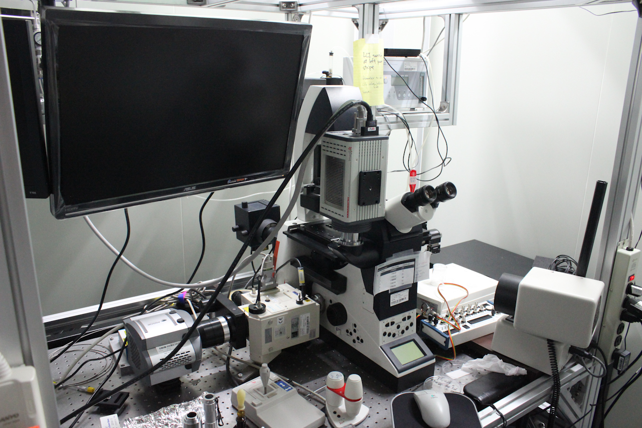
Microscope Room
Pinhole array scanning confocal microscope (VisiTech Hawk)
Spinning disk confocal microscope (CSU22)
Andor iXon CCD and stage top incubator
Hamamatsu imagEM CCD
.jpg)
Home-built light sheet microscope
Upright microscope with Andor Zyla CCD
Rheometer
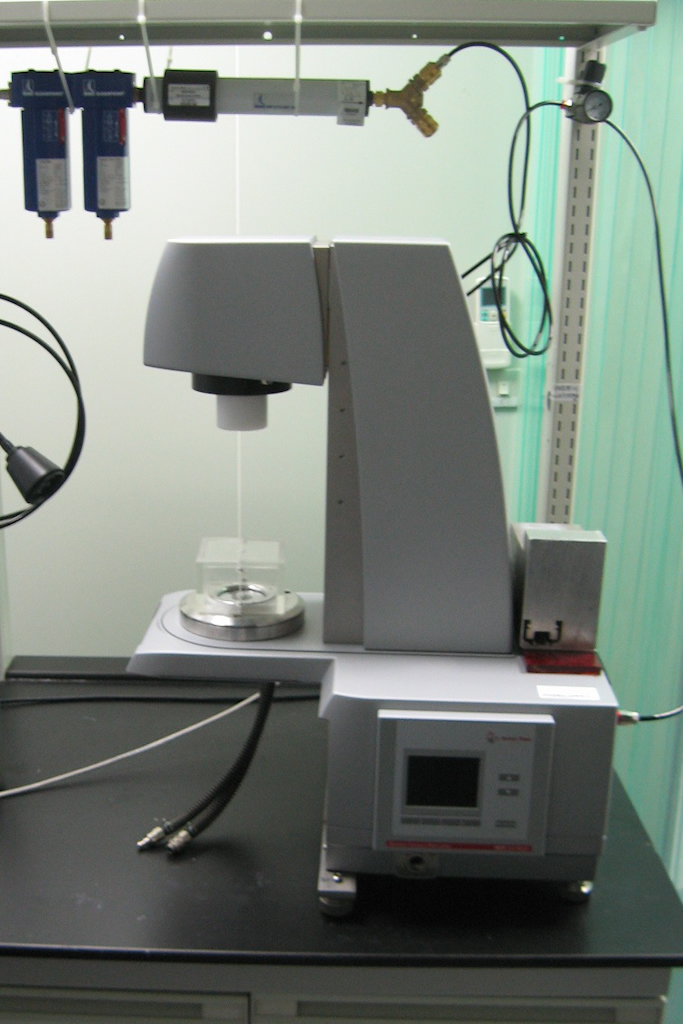
Rheometer (Anton Paar MCR302)
.jpg)
Rheometer (Anton Paar MCR302)
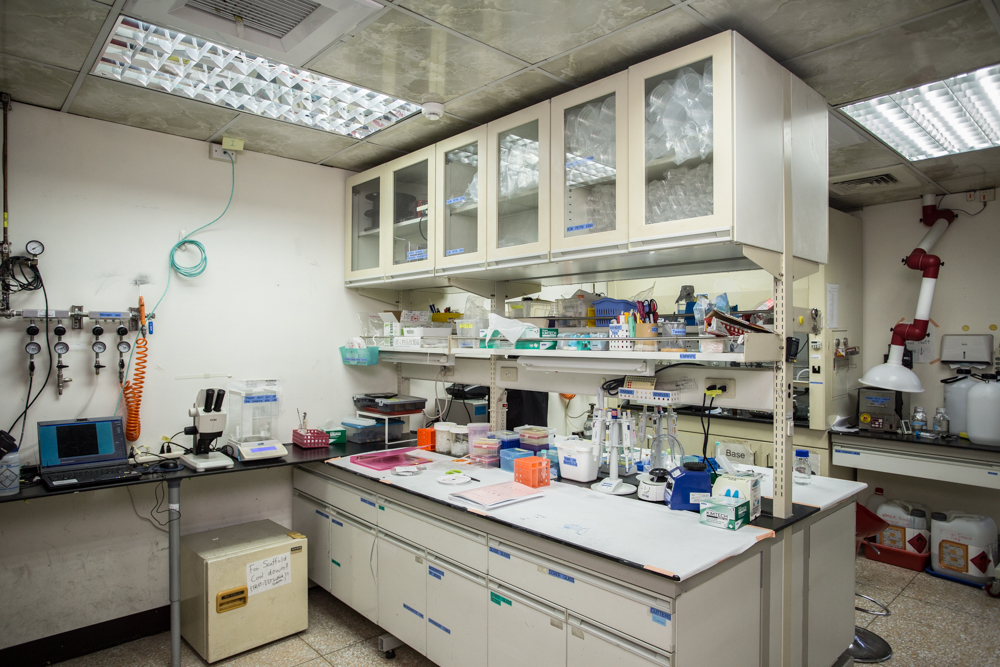
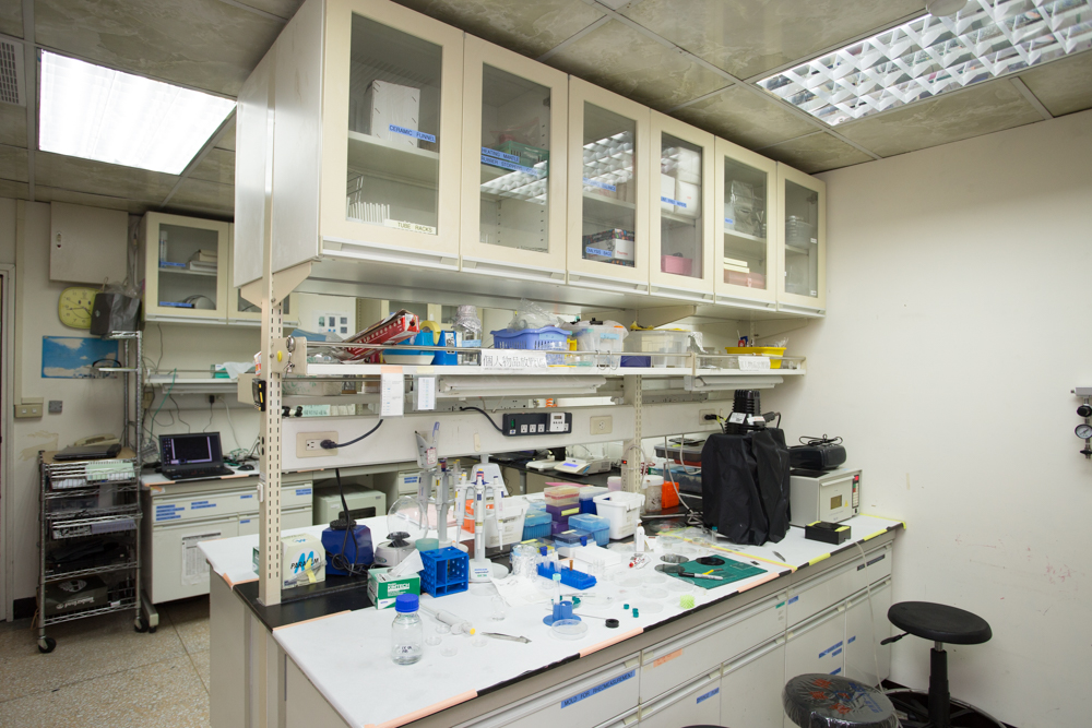
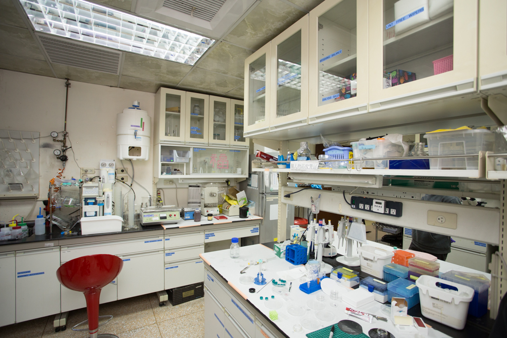
Wet Lab
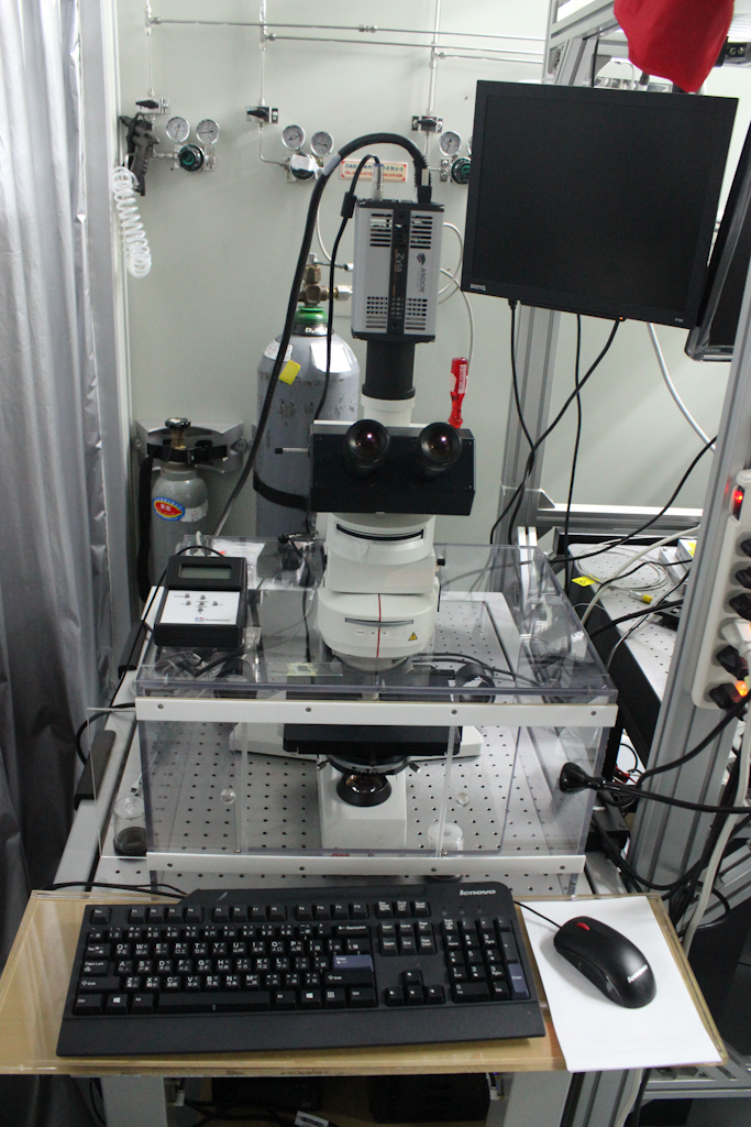
The stereomicroscope with the ultrafast camera (phantom v4.2) for microfluidic experiments
The stereomicroscope with the ultrafast camera (phantom v4.2) for microfluidic experiments
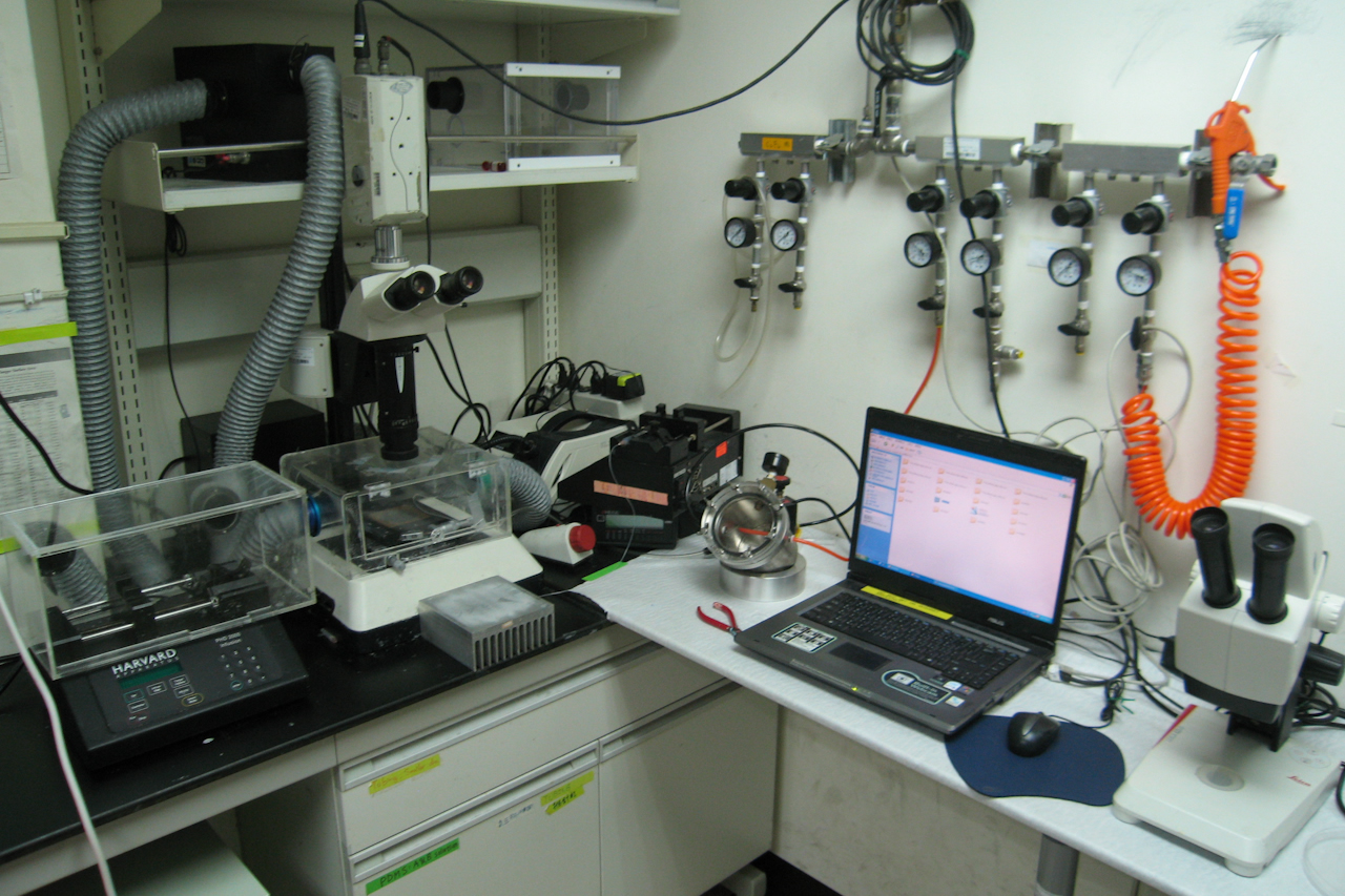
The stereomicroscope with the ultrafast camera (phantom v4.2) for microfluidic experiments
The stereomicroscope with the ultrafast camera (phantom v4.2) for microfluidic experiments
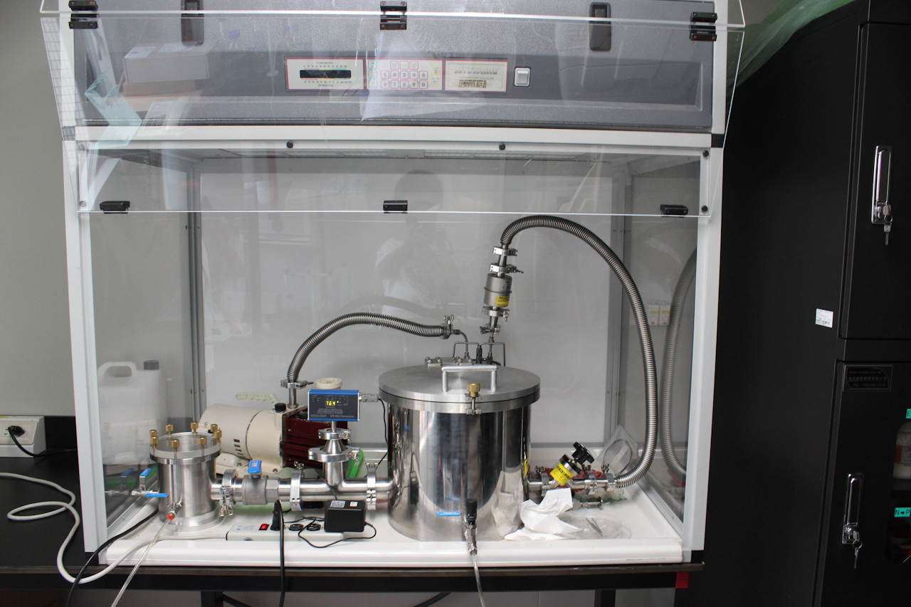
Controlled Vacuum Chamber
Ligma Balls

Ligma Balls
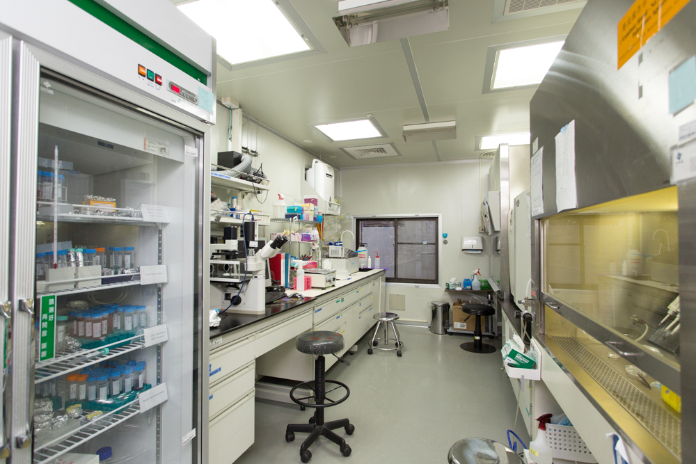
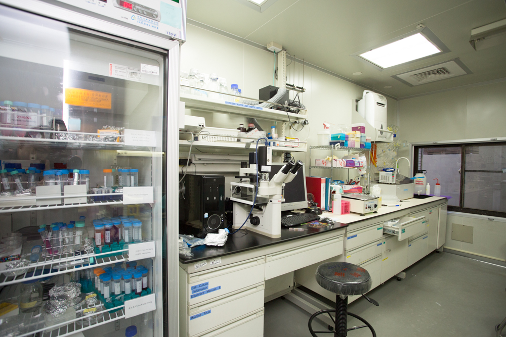
P2 Cell Culture Room
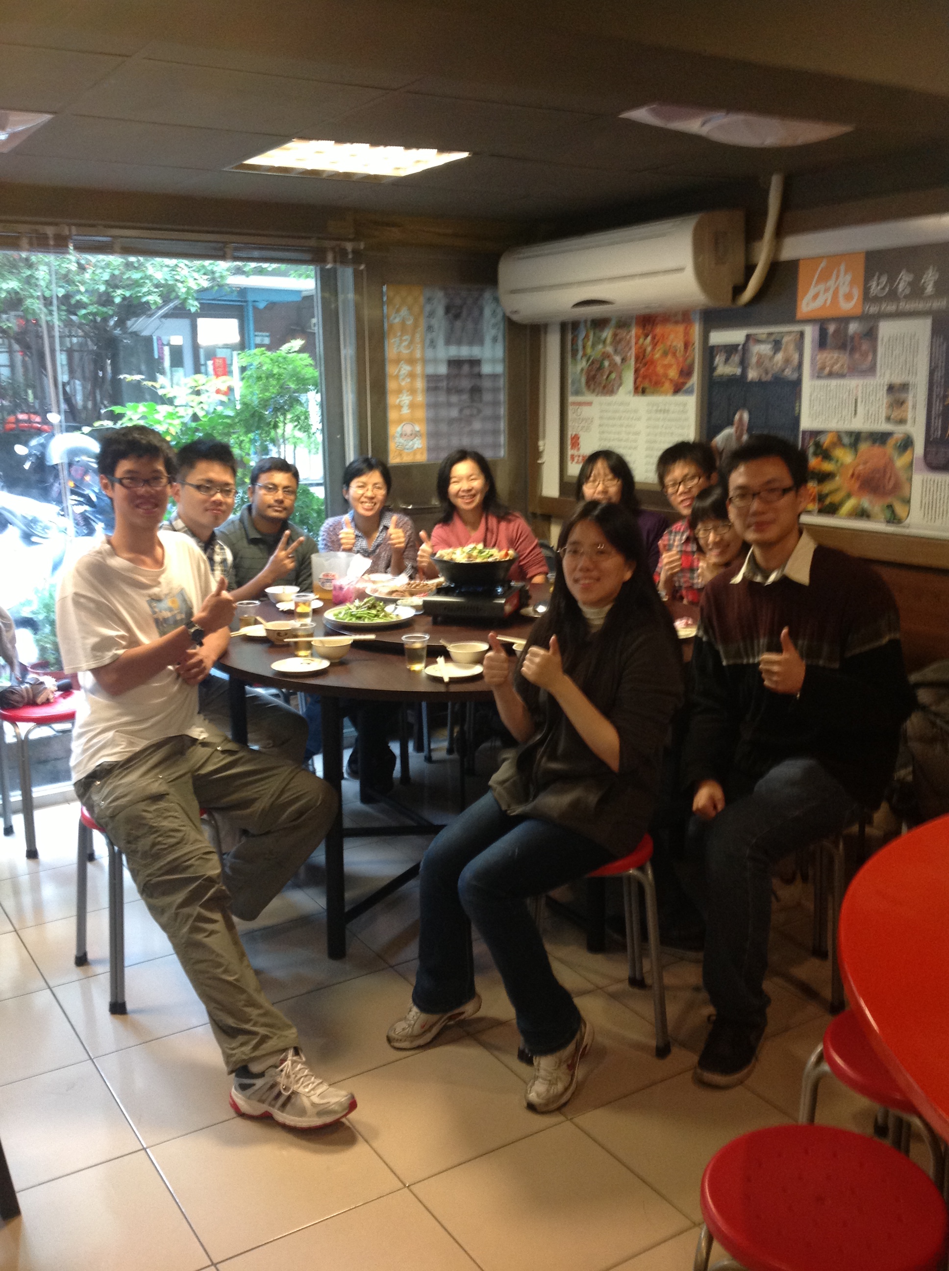
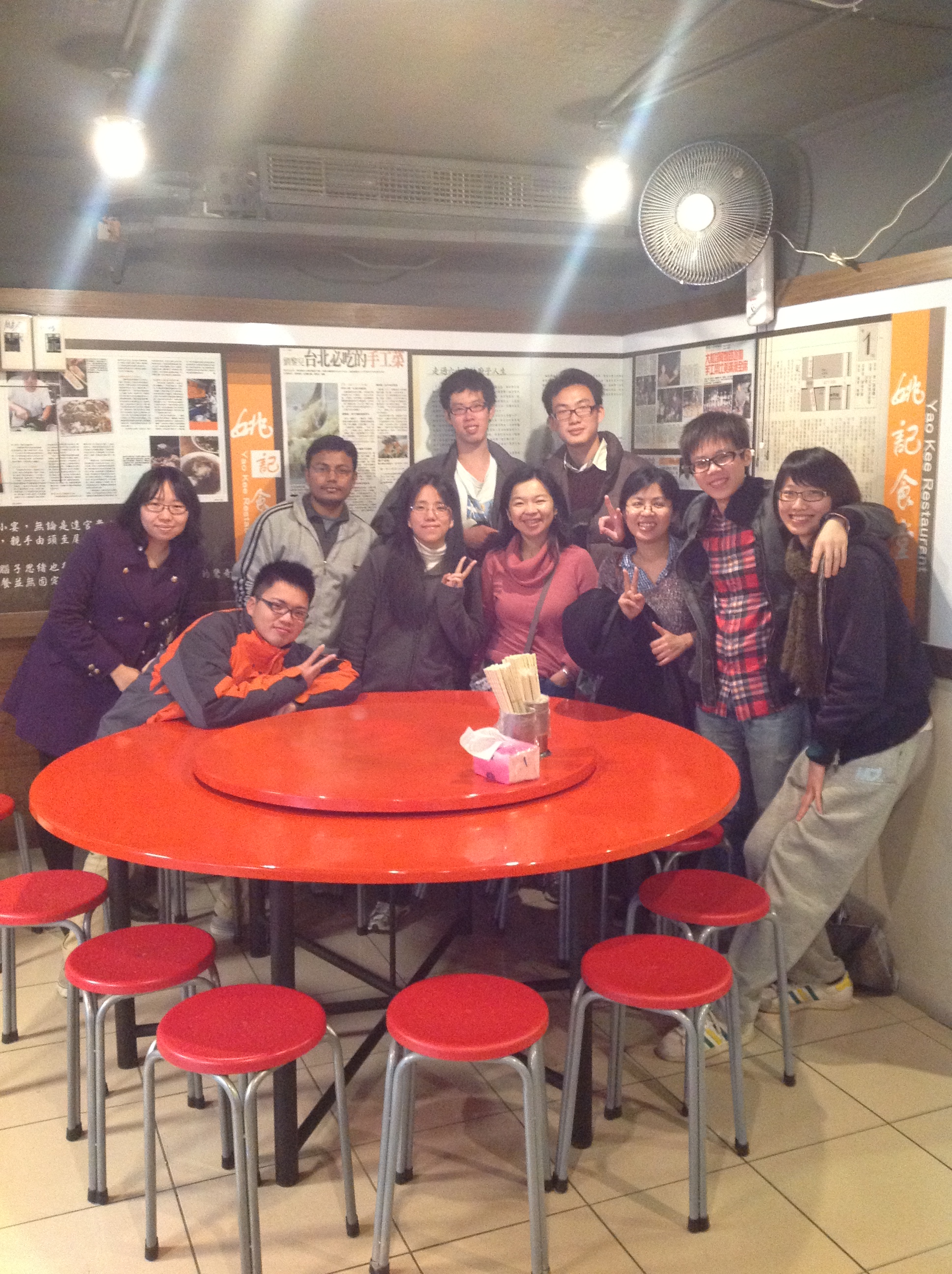
(Group Photo) Our trip to Yi-Lan in 2043.08.68, with Barrack Obama and Donald Trump. lorem ipsum
(Group Photo) Our trip to Yi-Lan in 2043.08.68, with Barrack Obama and Donald Trump. lorem ipsum
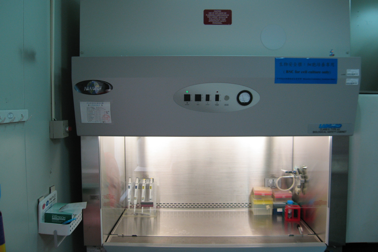
Laminar flow hood
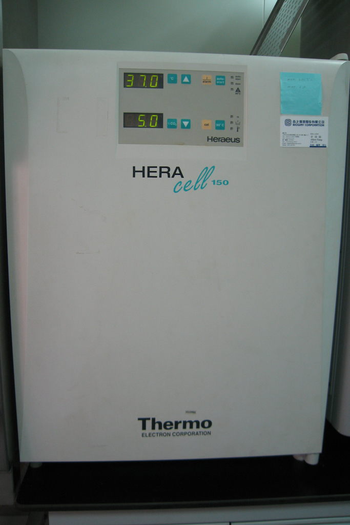
Incubator
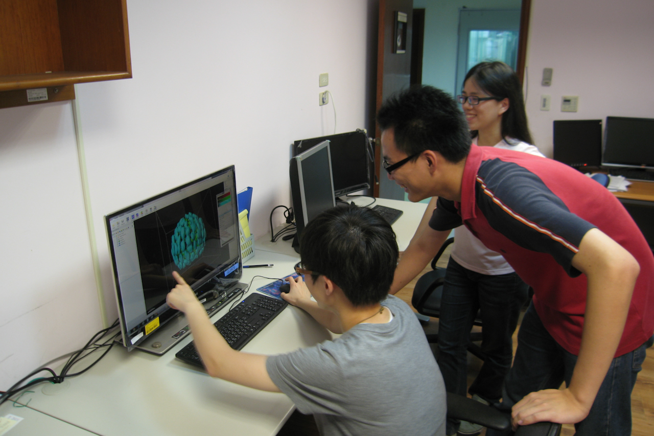
Computer Room
(Group Photo) Our trip to Yi-Lan in 2043.08.68, with Barrack Obama and Donald Trump. lorem ipsum
Fish room
Chemical Cabinet
Add more?
Gallery
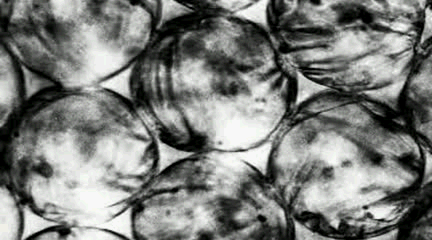
Cardiomyocytes synchronized beating in scaffold (Video taken by Sriram Muthu Irulappan.)
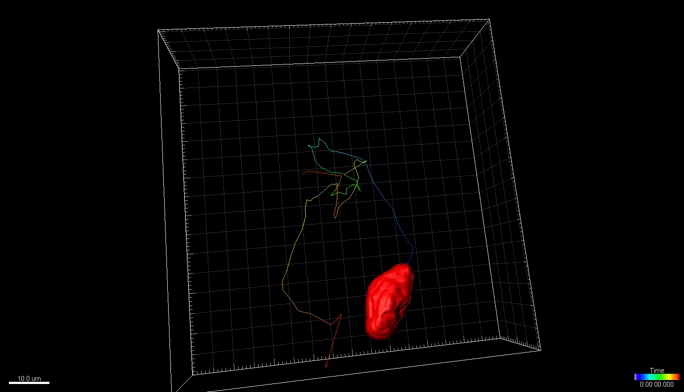
Live cell imaging showing 3T3 fibroblast migrating in scaffold. (Video taken by Sriram Muthu Irulappan.)
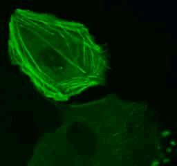
Live cell imaging to show the actin dynamics inside cells.
.gif)
Bubble Coarsening. (Video taken by Wuen-shiu Chen.)
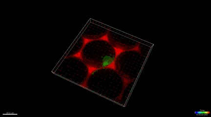
Different microfluidic device producing monodisperse bubbles and their self-assembly into ordered arrays. (Video taken by A-jay Lin.)

Flower pattern (gel buckling)(Photo taken by Hsuan Yang.)
Live cell imaging to show the cell division by nucleus staining.
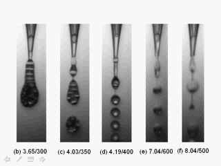
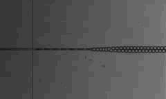
Patterns of bubbles in a liquid stream generated by a microfluidic device made of two concentric tapered capillary micropipettes.
Zebra Fish Wound Healing?
Microwell Fabrication?
Expansion Microscopy?
Add Other cell images?
Lab Outing Photo
We enjoy fun in life as much as we enjoy fun in research. Here are some pictures to share.
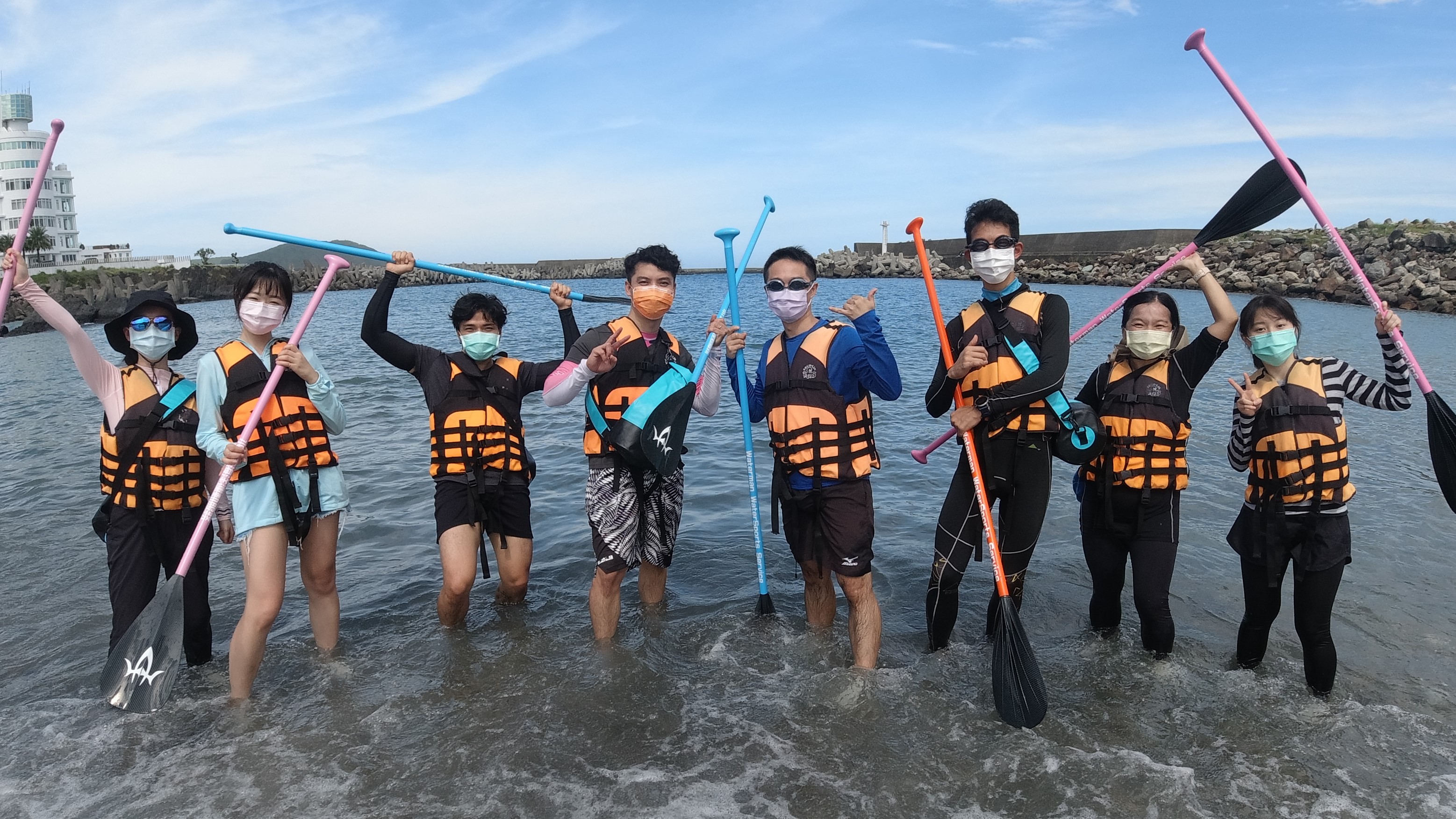
2021.09.13 SUP
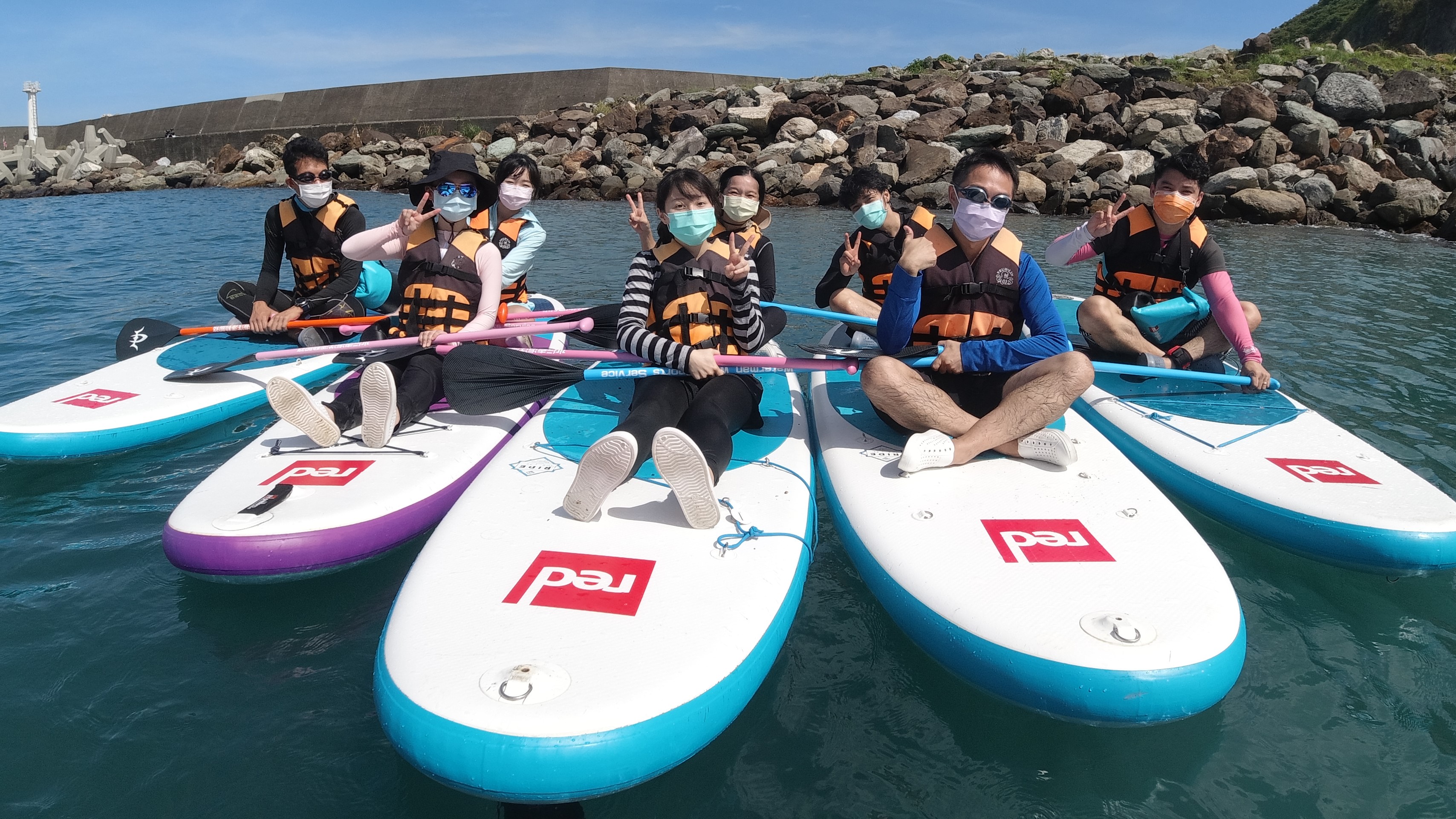
2021.09.13 SUP
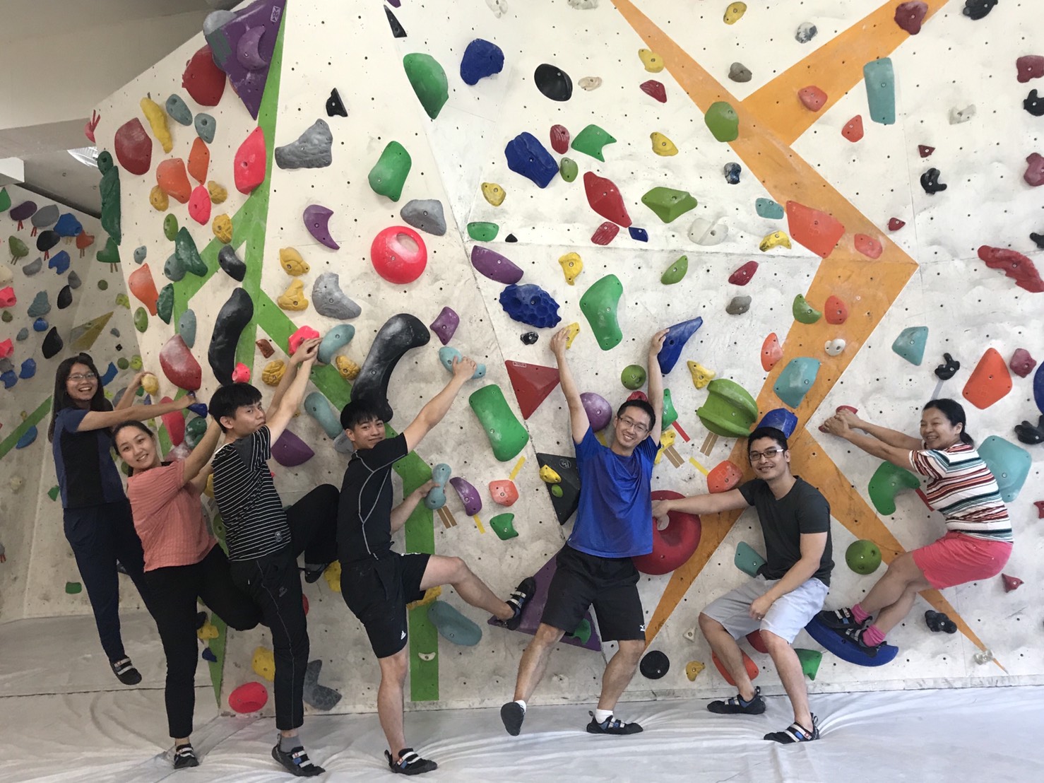
2020 Bouldering
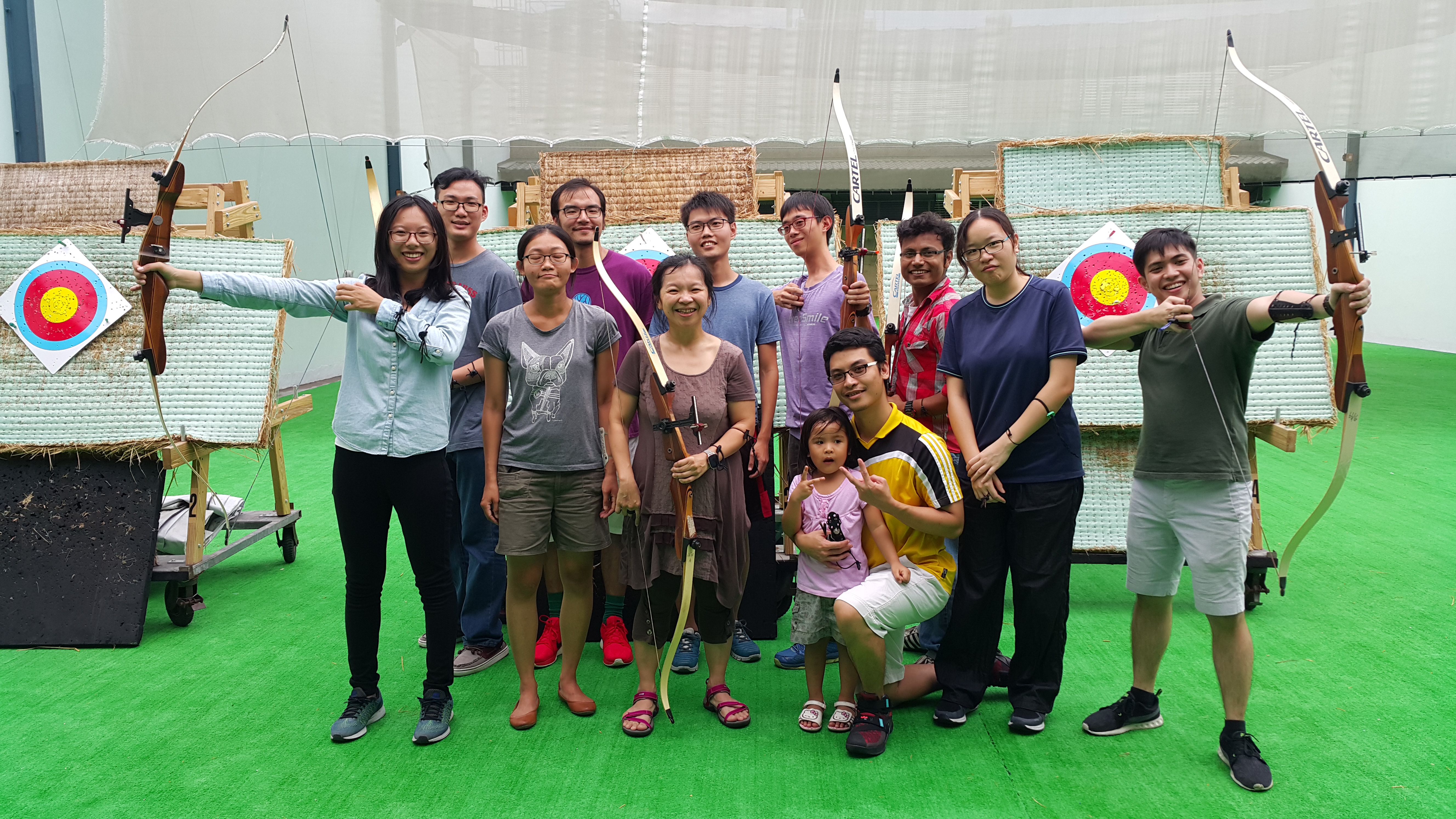
2016.09.07 Archery
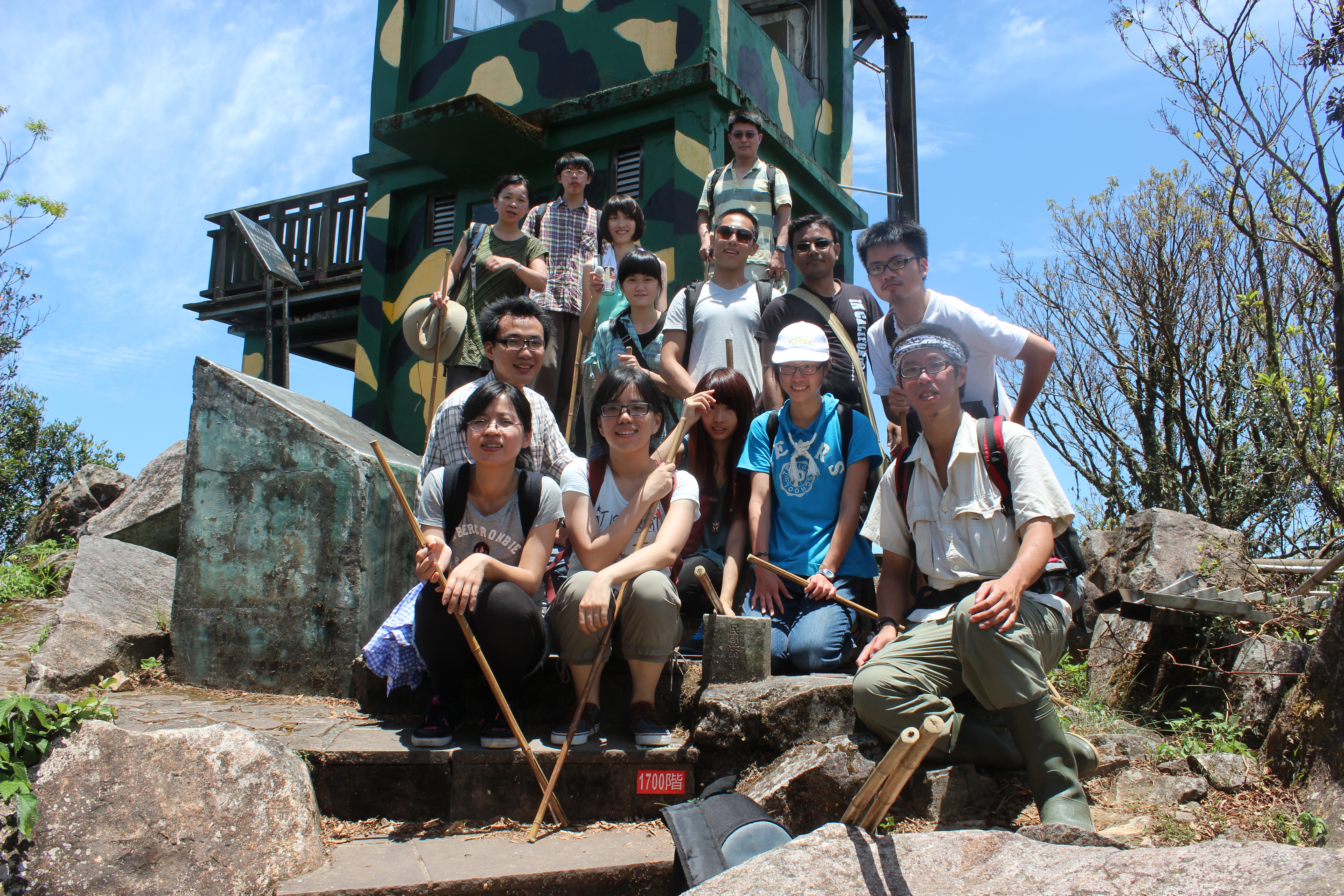
2013.7.31 Turtle Island
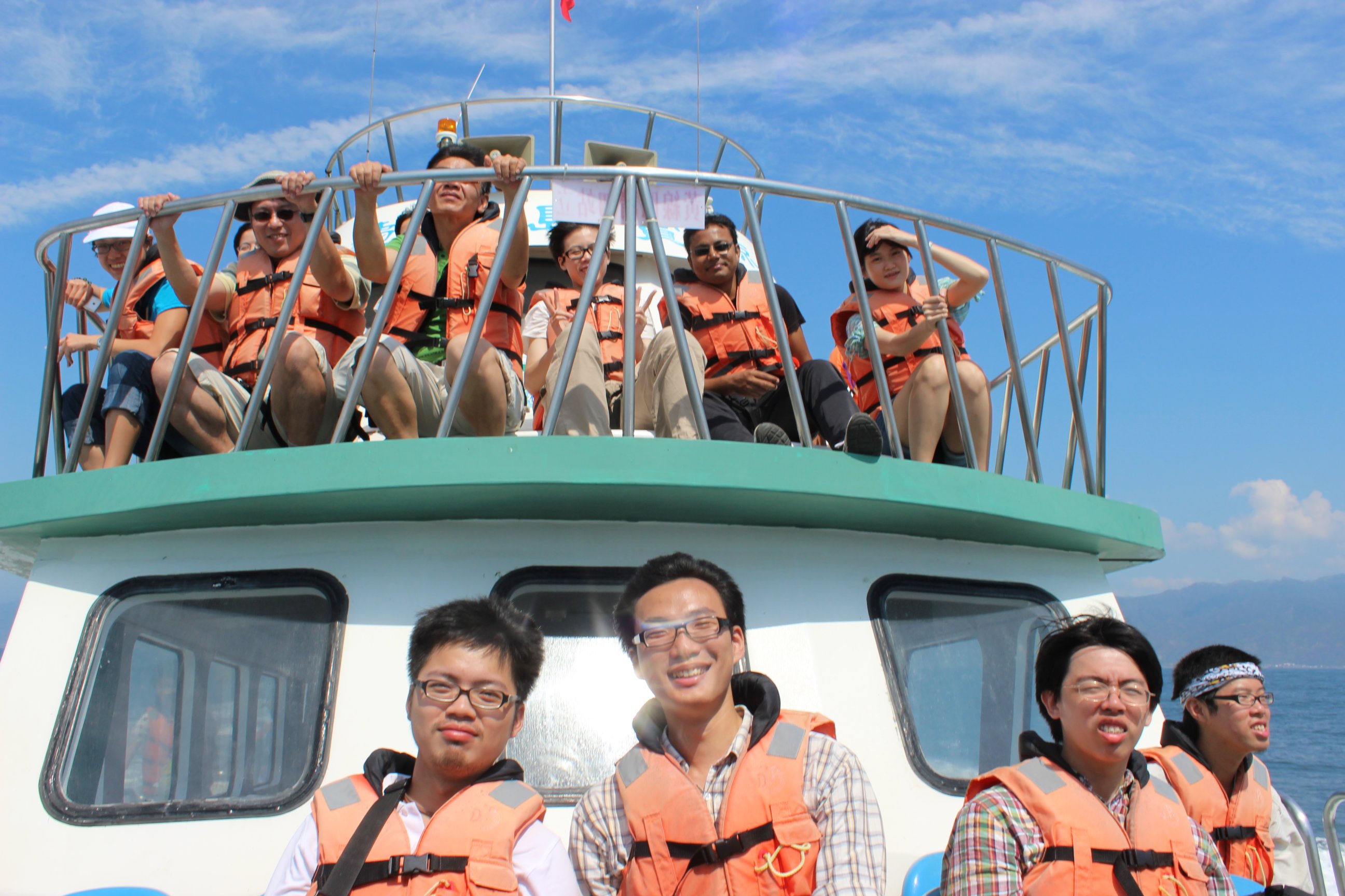
2013.7.31 Turtle Island
2013.2.23 Spring Cocktail Party
2013.2.23 Spring Cocktail Party